

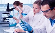
Pathology
Doctors
About the Department
The department started in 2010 along with the inauguration of Malabar Medical College and research centre, and is located at 7th floor in the academic complex. We provide excellent teaching services and comprehensive diagnostic services that helps clinicians in proper patient management.
The Pathology department has a 13 member faculty, assisted by 17 technicians and 7 clerical staff.
The various sections under this department are Histopathology, Cytology, Clinical pathology & Haematology and Blood bank. All the labs are equipped with skilled personnel and high quality calibrated instruments.
This department participates in EQAS programme on regular basis EQAS CBC- Biorad and EQAS coagulation in Haematology with CMC , Vellore. We routinely perform internal quality control checks also.
Departmental Focus
- To ensure good quality teaching services to undergraduate students of various faculties like MBBS, BDS, Nursing and Paramedical sciences.
- To incorporate innovative teaching methodologies into students’ curriculum.
- To provide state of the art diagnostic services which would help in patient management. We intend to start advanced diagnostic techniques like Immunohistochemistry (IHC) in the near future.
Infrastructure
HISTOPATHOLOGY
Our histopathology lab is located on the ground floor with easy access to patients. Lab issues reports roughly on 7000 biopsy specimens a year.
- HPE (small biopsies like endoscopic mucosal biopsies, curettings, punch biopsies of oral cavity, cervix, trucut biopsy from breast, prostate and small incisional biopsies).
- HPE (big biopsies like hysterectomy, thyroidectomy mastectomy, gastrectomy, , total colectomy, and other organs.
- Decalcification and processing for bone biopsies
Routine tests:
- Histochemistry – Periodic Acid Schiff (PAS), Giemsa, Perl’s stain
- Immunohistochemistry
Special tests:
CYTOLOGY
With a dedicated team of pathologists, diagnosis is reached for materials obtained by fine needle aspiration and exfoliation. The department provides reports approximately on 6500 cytology specimens in a year.
- FNAC (Fine needle aspiration cytology) – Procedure andreporting
- Exfoliative cytology including PAP smears, body fluids, sputum, bladder and bronchial washings
- Cell block studies
Tests done:
HAEMATOLOGY AND CLINICAL PATHOLOGY
Our haematology and clinical pathology section which is located in the central lab (ground floor) is equipped with one 3 part and one 5 part automated cell counters (Mindray). We offer prompt services round the clock. Lab also provides facilities for coagulation studies using
On an average the lab provides reports on around 5000 peripheral smears, 3 lakh haematology tests and 2,40,000 clinical Pathology investigations a year.
We ensure good quality and accurate reporting services as judged by enrolling in regular IQAS and EQAS programmes.
- Complete blood count
- ESR
- Peripheral smear
- Coagulation studies – Bleeding time, clotting time, PT, APTT
- Urine routine examination.
- CSF and other fluid cell counts.
- Semen analysis
- Mantoux test
- Urine pregnancy test
- Stool occult blood
Tests done:
BLOOD BANK
Blood banking services have been provided by MMCH since the start of the institution. The blood bank is equipped with most modern calibrated equipments (Terumopenpol) and well trained technicians. Our blood bank has evolved into a full fledged unit since the installation of component separation unit in January 2017. We have been able to meet the needs of patients for the different blood components since then. Currently we are also issuing blood units to needy patients at other hospitals. We have introduced the gel card system from TULIP for blood grouping and cross matching since January 2017. Our Blood bank functions in accordance with the regulations laid down by Drugs control department and issues monthly report to KSACS.
- Blood grouping and cross matching
- Screening for transfusion transmissible diseases
- Component separation – Blood components available are PackedRBC,PRP,Platelet concentrate, FFP, Cryoprecipitate.
Procedures done:
TEACHING FACILITIES
- Two demonstration class- capacity of 75 each
- Museum- 400 mounted specimen and 50 wet specimen
The department conducts theory and practical classes on a routine basis for undergraduate students in different faculties. The teaching faculty are well qualified and take keen interest in introducing innovative methods of teaching. We also have an excellent museum which displays around 400 biopsy specimens.
Publications
1. Primary Meningeal Melanocytoma, Kathmandu UniversityMedical Journal(2008),vol6,No. 2,Issue 22,245-247.
2. Papillary carcinoma Thyroid, A 11year epidemiological study with histopathological correlation in a tertiary care centre in south Malabar region in Kerala. India. Journal of Pathology of Nepal. Sept 2015 vol5,no2,issue 10,798-805
3. An autopsy study of heart with an emphasis on coronaries.Annals MedicusVol2,issue02,April 2015.
4. SilySreedharan, Prema N S Pseudohirchsprung’s disease- a clinicopathological correlative study .Indian Journal of Pathology and Oncology, April-June 2015;2(2);69-72
5. SilySreedharan&Prema N. S.Hirchsprung’s Disease -A Clinicopathological Correlative Study International Journal of Current Medical And Applied Sciences, 2015, August, 7(3),155-160.
6. SreedharanSily, Nair Geethu G Cytomorphology of
recurrent back swelling-eccrinespiradenoma Indian Journal
of Pathology and OncologyYear : 2015, Volume : 2, Issue : 2
7. SreedharanSily, RaoVineeth, Thomas Malini. Diffuse intestinal ganglioneuromatosis with coexisting gastrointestinal stromal tumour - gastrointestinal manifestations of neurofibromatosis: an unusual case report. International Journal of Research in Medical Sciences. 2015 Sept; 3(9): 2499-2502.
8. Shamsuddin F, Ilias LM, Sreedharan S. A comparison of didactic lectures to self-directed learning in medical education. J. Evid. Based Med. Healthc. 2017; 4(3), 110- 114. DOI: 10.18410/jebmh/2017/22.
9. Shamsuddin F, Khadilkar UN, Saha D, Sreedharan S. A clinicopathologic study of mediastinal lesions with special emphasis on thymomas. International Journal of Research in Medical Sciences 2015;3(8):1902-10.
10.Shamsuddin F, Khadilkar UN, Saha D, Sreedharan S. Mediastinal lymphomas – A clinicopathologic study. Indian Journal of Pathology and Oncology 2015;2(3):131-140.
11.Shamsuddin F, Khadilkar UN, Saha D. Unusual lesions of the mediastinum. Lung India 2015;32:566-71
12.Comparison of didactic lectures to self directed learning in Medical education – Dr Fatima Shamssudin. (Published in Journal of evidence based Medicine;Jan 2017; 4(3); 2349-2570.
13.Implementation of Bethesda system for reporting thyroid cytology in an academic institution: A 2 year retrospective study Sily Sreedharan , Deeshma , Fatima Shamsuddin , Chris Thomas , Shobhitha D IP Journal of Diagnostic Pathology and Oncology, April-June, 2018;3(2):127-132
CURRENT RESEARCH
- Comparison of Pocket Chem –Hemo G method with Cyanmethhaemoglobin method and automated cell counter method for estimation of haemoglobin in the age group between 18-70yrs in a Private medical college setting- Dr Sily Sreedharan ,Dr Deeshma T, Dr Fatima Shamsuddin.
- Histopathological and Immunohistochemical Analysis of GIST
Other Activities
- Department conducts Pathology quiz for undergraduate students in December every year after completion of course.
- Department encourages students to participate in quiz conducted outside the college too.
- Ramachandran memorial pathology gold medal exams are conducted every year in January . Student have secured 4rth place in 2016 gold medal exam.